0441
Improved spiral dynamic MRI of vocal tract shaping at 3 Tesla using dynamic off resonance artifact correction1Biomedical Engineering, University of Iowa, Iowa city, IA, United States, 2Ming Hsieh Department of Electrical Engineering, University of Southern California, Los Angeles, CA, United States, 3Department of Radiology, University of Iowa, Iowa city, IA, United States
Synopsis
Spiral trajectories have been valuable in several dynamic speech production MRI studies due to its robust motion encoding, and time acquisition efficiency. However they are challenged by off-resonance artifacts at high field strengths and use of long readouts. In this work, we demonstrate correction of off-resonance induced blurring and signal losses on 3T spiral dynamic speech MRI data. The approach estimates the dynamic field map directly from the single TE data and compensates for this during a multi-frequency interpolation reconstruction. Effectiveness in improving sharpness of various articulator boundaries for long readout sequences (> 2ms) are demonstrated.
Purpose
Spiral trajectories have been valuable in several dynamic speech production MRI studies due to its robust motion encoding, and time acquisition efficiency. A known limitation is its sensitivity to off-resonance artifacts, which can manifest as signal loss/blurring artifacts at various air-tissue interfaces (eg. at the lips, tongue, velum) [1-2]. This effect is more pronounced at high field strengths (>3 T) and with the use of long readouts. Radial trajectories therefore have been commonly used at 3 T due to their short readouts, and robustness to off-resonance despite their lower acquisition efficiency compared to spirals [3-4]. Several de-blurring methods exists that rely on dual TE acquisitions to estimate the time-varying field map [5], or employ non-convex auto-focus metrics [6-7]. Recently, a single TE based dynamic off-resonance correction scheme has shown to be effective to improve articulator sharpness for long readout spirals (upto 5 ms) at 1.5 T [8]. In this work, we evaluate the utility of this scheme on spiral speech data with varying readout lengths at 3 T.Methods
Following the approach in [8], we estimate the dynamic field map from the phase of coil combined time series image data after performing a coil phase compensation as . The complex coil maps are estimated from the data itself using the sum of squares approach from time averaged image data after spatial low-pass filtering with a 2D Hanning window with size 21x21. is then obtained using optimal B1 coil combination. At 3T, off-resonance at the air-tissue interface can be up to ~1200Hz, which leads to phase wrapping in . We additionally perform phase unwrapping [9] on prior to field map estimation. This issue could alternately be suppressed by using a shorter TE. Note, the above approach assumes the coil maps are spatially smooth and stationary, and therefore the time varying field map can be directly estimated from .
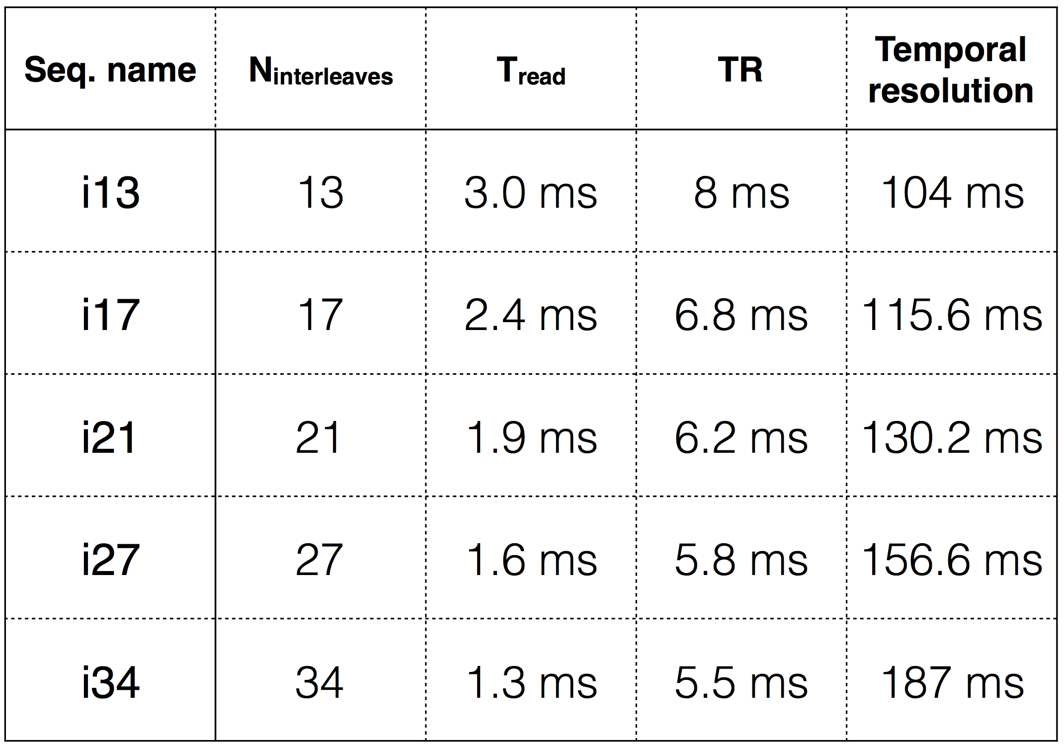
Experiments were performed on a GE-750 W 3T scanner with a 16 channel head and neck coil. 6 channels that were most sensitive to the vocal tract regions of interest (eg. lips, tongue, velum) were used. Various multi-shot variable density spiral (VDS) free induction decay (FID) sequences were realized with different choices of interleaves (Nintl) and therefore different read out times (Tread). Golden angle time interleaving was employed. Imaging parameters were: spatial resolution: 2.4x2.4 mm2; slice thickness=6 mm; flip angle = 150; TE=1.5 ms; FOV in VDS was linearly varied from 40x40 cm2 to 8x8 cm2 along the k-space radius. Other key sequence parameters are listed in figure 1. A 50 year old speaker was imaged who performed tasks of opening and closing the mouth, and repeated utterance of “loo-lee-la-za-na-za”. For every sequence, view sharing gridding reconstruction was performed onto a 84x84 matrix size with Nintl per frame and a time step of 3 interleaves using the multi-frequency interpolation method [10] based on the estimated dynamic field map. Image quality was qualitatively compared without and with dynamic off-resonance correction.
Results
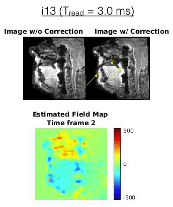
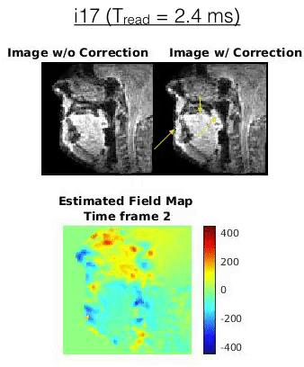
Figure 2 shows images where the subject had his mouth open. The sequences i13, and i17 showed considerable off-resonance signal loss (eg. at the lips, tongue tip; see red arrows), and blurring (eg. at the velum; see yellow arrows). These artifacts are reduced with off-resonance correction resulting in improved depiction of the articulator boundaries. Subtle gains in image sharpness were also observed in sequences i21, i27, and i34 (see green arrows). Figure 3,4 (animations) respectively shows animated time series of the images from i13 and i17 without and with correction when the subject was speaking loo-lee-la-za-na-za. It can be seen that the moving air-tissue interfaces results in time varying spatially dependent changes in the estimated field map. With off-resonance correction, the image quality is significantly improved in terms of reduced signal losses and improved sharpness. Note the spiral flickering artifacts in i17 are due to the choice of $$$Nint=17$$$ not being a fibonacci number which results in a nonuniform sampling in k-space.Conclusion
We demonstrated feasibility to correct for off-resonance induced artifacts in dynamic speech MRI for long readout (> 2 ms) spiral sequences at 3 T. The scheme estimates the dynamic field map directly from single TE data. Results from a single volunteer showed utility in restoring signal losses and improving sharpness at various articulator boundaries (particularly at the lips, tongue tip, and velum). One known limitation is that this approach only compensates for time varying field maps and does not compensate for a time averaged field map. Future work will include integration with sparsity based reconstruction, and quantitative validation on number of subjects.Acknowledgements
No acknowledgement found.References
[1] Lingala SG, Sutton BP, Miquel ME, Nayak KS. Recommendations for real-time speech MRI. J. Magn. Reson. Imaging 2016;43:28–44. doi: 10.1002/jmri.24997.
[2] Scott AD, Wylezinska M, Birch MJ, Miquel ME. Speech MRI: Morphology and function. Physica Medica. 2014;30(6):604–618.
[3] Niebergall, Aaron, et al. "Real‐time MRI of speaking at a resolution of 33 ms: Undersampled radial FLASH with nonlinear inverse reconstruction." Magnetic Resonance in Medicine 69.2 (2013): 477-485.
[4] Burdumy M, Traser L, Richter B, et al. Acceleration of MRI of the vocal tract provides additional insight into articulator modifications. J Magn Reson Imaging. 2015
[5] Sutton BP, Conway CA, Bae Y, Seethamraju R, Kuehn DP. Faster dynamic imaging of speech with field inhomogeneity corrected spiral fast low angle shot (FLASH) at 3 T. J Magn Reson Imaging. 2010;32:1228-1237.
[6] Chen W, Meyer CH. Fast automatic linear off-resonance correction method for spiral imaging. Magn Reson Med. 2006;56:457- 462.
[7] Smith TB, Nayak KS. Automatic off-resonance correction in spiral imaging with piecewise linear autofocus. Magn Reson Med. 2013;69:82-90
[8] Lim Y, Lingala SG, Narayanan SS, Nayak KS. Dynamic off‐resonance correction for spiral real‐time MRI of speech. Magn Reson Med 2018. doi:https://doi.org/10.1002/mrm.27373
[9] M. A. Schofield and Y. Zhu, Fast phase unwrapping algorithm for interferometric applications, Opt. Lett., vol. 28, no. 14, p. 1194, 2003
[10] Man LC, Pauly JM, Macovski A. Multi frequency interpolation for fast off-resonance correction. Magn Reson Med 1997; 37:785-792
Figures